How Does Your Baby Grow – Photos
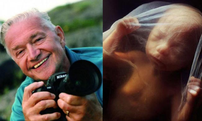
Photographer Lennart Nilsson has spent 10 years of his life to picture the evolution of the embryo from conception to birth.

Nilsson received international acclaim in 1965, when the LIFE magazine published 16 pages of the photos of a human embryo. These photos were immediately replicated in Stern, Paris Match, The Sunday Times and other magazines, bringing world fame to photographer Lennart Nilsson.
Since childhood, microscope and camera have been Nilsson’s main hobbies, as he wanted to share with the world the beauty of the human body at the cellular level. Nilsson managed to get the pictures of the human embryo back in 1957, but they were not spectacular enough to show them to the public.
The most accurate and colorful pictures were acquired with the help of a cystoscope – a medical device used to examine the bladder from the inside. Nilsson attached a camera and an optical fiber to it and made thousands of pictures of the baby’s life inside the womb.
Lennart Nilsson’s skillful hands have created a miracle and familiarized the world with the mystery of the origin of human life.

The sperm cell moves in the fallopian tube toward the egg cell.

An egg cell.

A fateful encounter.

One of the father’s 200,000,000 sperm cells has broken through the egg shell.

The sperm cell in the section. Its head contains the entire genetic material.

A week later, the embryo is sliding down the fallopian tube, moving to the uterus.

A week later, the embryo is attached to the uterine mucosa.

The 22nd day of the embryo development. The gray matter is the future brain.

On the 18th day, the embryo’s heart beating starts.

The 28th day after fertilization.

5 weeks. The length is 9 mm. The face can be discerned, as well as the openings for the mouth, nostrils and eyes.

40 days. The outer cells of the embryo are fused with the loose surface of the uterus and form a placenta.

8 weeks.

10 weeks. The eyelids are half-open. Within a few days, they will form completely.

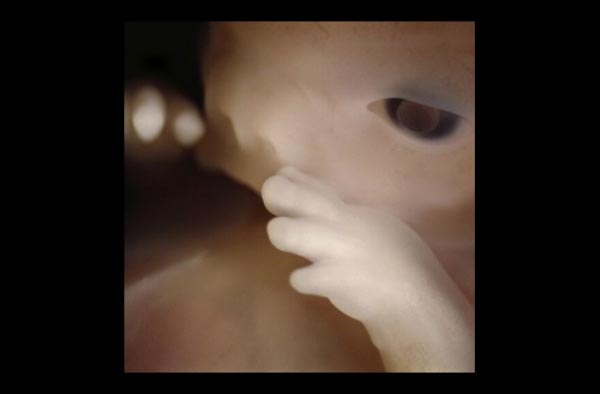
10 weeks. The baby is already using its hands to explore the area.

16 weeks.

A network of blood vessels is visible through the thin skin.

18 weeks. The fetus can perceive sounds from the outside world.

19 weeks.

20 weeks. The height is about 20 cm, and hair is emerging on the head.

24 weeks.

6 months.

36 weeks. In a month, the baby will be born.
In 1965, Nilsson published a book of photographs called “A Child is Born”, and it was sold out in a few days. The book has many editions and still remains one of the bestselling books in the history of this kind of photo albums.
The pioneer of medical photography Lennart Nilsson is 91 years old. He is still involved in science and photography.
